The cells in Daolin Tang’s Petri dishes were dead. Signs of life had dropped off a cliff; they weren’t reproducing. They were goners. And that was good; the cell biologist had been looking for chemicals that would poison cancer cells. These secret battles between your body’s cells might just save your life But Tang and
The cells in Daolin Tang’s Petri dishes were dead. Signs of life had dropped off a cliff; they weren’t reproducing. They were goners.
And that was good; the cell biologist had been looking for chemicals that would poison cancer cells.
These secret battles between your body’s cells might just save your life
But Tang and his colleagues faced a post-mortem mystery: the precise cause of death. They investigated several ways in which cells can die, and none of them was guilty.
Eventually, the researchers determined that the dying cells had an unusually high internal pH. They died because they were basic.
The team named the phenomenon alkaliptosis1. The work not only expanded scientists’ understanding of how cells can die and how pH controls their fate, but also suggested an entirely new way to kill cancer cells, says Tang, who did the studies while at the University of Pittsburgh in Pennsylvania and now works at the University of Texas Southwestern Medical Center in Dallas.
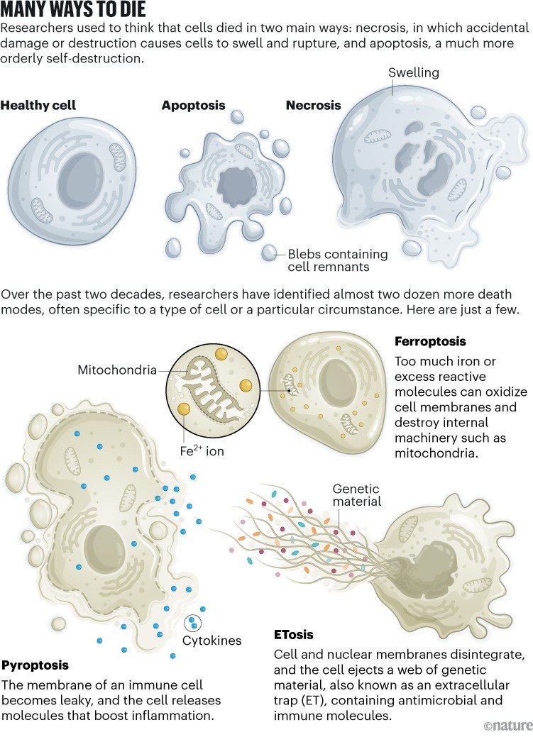
Tang’s experience has been repeated again and again in the twenty-first century, with scientists describing 20 or so new kinds of cell death — and counting — since 1999 (see ‘Many ways to die’). This is a major change: researchers used to think there were two main ways cells could die. Death could happen by accident, such as when a cell is squashed, sliced or otherwise destroyed; this is called necrosis. The other classical option is apoptosis: a highly regulated, stepwise and tidy process in which the cell releases little ‘blebs’ of membrane that the immune system can sweep away.
For a long time, those two options stood alone. Perhaps that’s because cell death is hard to study. “If you have a successful cell-death experiment, your material disappears,” jokes Scott Dixon, a cell biologist at Stanford University in California.
But the advent of modern scientific technologies, such as gene editing, high-resolution imaging and diverse ‘omics tools, has revealed many fresh forms, says Xingbin Hu, a cell biologist at Xijing Hospital in Shaanxi, China. There’s fiery, pro-inflammatory pyroptosis; explosive, immune-activating necroptosis; and iron-induced, membrane-destroying ferroptosis. At least four new types were defined in 2025 alone. This spring, scientists studying flatworms described another, called ruptosis, in which particular cells explode to splatter toxins over invading bacteria2. Cells might even have mechanisms to delay their own death, or to pull back from the brink and live on.
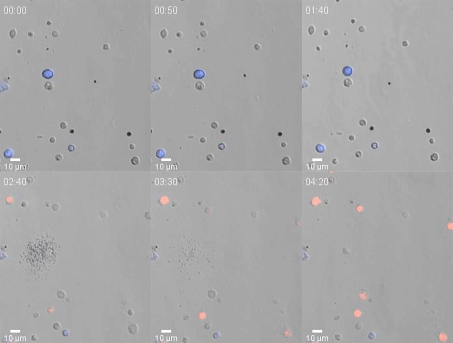
Cells called ruptoblasts, discovered this year, can explode and splatter immune agents that kill invaders.Credit: Dr Eliya Sultan
The manner in which cells perish matters, says Tang. Dying cells release signals or material that affects neighbouring cells, shapes tissues and their functions and influences immune responses and disease. For example, after being invaded by a pathogen, a cell can die by suicide, but as it dies, it might leak its contents — which calls in immune cells to fight the broader infection. So understanding how cells die, or don’t when they should, has ramifications for researchers’ understanding of development, metabolism and immunity.
Scientists are linking these newfound death pathways to diseases and eagerly investigating medical applications, such as killing cancer cells, calming inflammatory disease and even rescuing dying cells in the heart and brain.
The discovery of these new death modes highlights how much we still have left to learn about basic biology, says Tang. “Cell death is not simply the end of a cell’s life.”
Into oblivion
Nor is it rare: every day, billions of cells in the human body shuffle off this mortal coil. Damaged, used up or simply no longer needed, they wither, harden or explode. “All the cells in creatures must know when, how and where to die,” says Hu. “With so many different cells playing unique roles in life, it’s not strange that various cell death forms are quite needed.”
Too much of a compound can kill a cell, and death will look different depending on the source; for example, researchers described death by excess copper3 in 2022 and by sodium overload4 in 2025. These findings reveal the limits of cellular survival, says Qing Zhong, a biochemist at the Shanghai Jiaotong University School of Medicine in China, who led the work on sodium-based death.
Cells have a menu of self-inflicted death modes, such as apoptosis and pyroptosis. If all goes well, these leave the organism better off. During development, some cells, such as those that give human embryos webbed fingers, undergo apoptosis because their job is done. Others, such as those lining the gut, have a finite lifespan, and make way for fresh versions of themselves. Some skin cells, with a death mode called cornification, leave their corpses behind to serve as the body’s outermost, protective layer.
Infected cells often sacrifice themselves for the greater good of the organism, to stymie viruses or bacteria that would use them to reproduce. Infection was probably a driver of the evolution of several death modes, because infected cells needed backup options to ensure that they could dispatch themselves, speculates Ana García-Sáez, a biophysicist at the Max Planck Institute of Biophysics in Frankfurt, Germany. Some viruses can inhibit individual death pathways to keep host cells alive; if the cell could switch to another death programme, such as pyroptosis or necroptosis, it could still thwart the virus. “It’s like a race with the pathogen,” García-Sáez says.
From the jaws of death
Scientists have found another kind of flexibility: cells don’t always proceed directly from a death trigger to their demise. They might spend time in limbo, or even survive.
Take gut epithelial cells. Dying cells usually excise themselves from the intestinal lining, taking an active role in their own disposal. But that process takes time, and the cells need to live long enough to complete it. A specialized enzyme helps them to hang on while they do this, as described in a 2022 paper5 by immunologist Edward Miao at Duke University in Durham, North Carolina, and his then-postdoc Kengo Nozaki — now an immunologist at the Medical University of South Carolina in Charleston.
They took mice missing the enzyme caspase-7, which facilitates cell death, and infected them with Salmonella, bacteria that cause food poisoning. Infected gut epithelial cells began to die. But without caspase-7, they stuck around instead of exiting the gut lining, causing extra inflammation and tissue damage. Their death throes cause holes to form in the cell membrane, and Nozaki and Miao found that caspase-7 usually activates repair of those holes so that dying cells can survive long enough to make a clean getaway.
Nozaki and Miao proposed in a 2023 review that many cells have a “bucket list” of tasks to complete before they expire6. Some immune cells use these tasks to protect the body from pathogens. A macrophage infected with bacteria can die by pyroptosis, but in doing so it releases distress signals and arranges its cellular membranes to trap the pathogens in its corpse7. And neutrophils, which live for only hours to days, might take drastic, offensive action on the way out. Should they encounter bacteria, they can unwind their DNA, dissolve their nuclear membrane and burst. As they explode, they release a web made of their own genetic material to ensnare invaders.
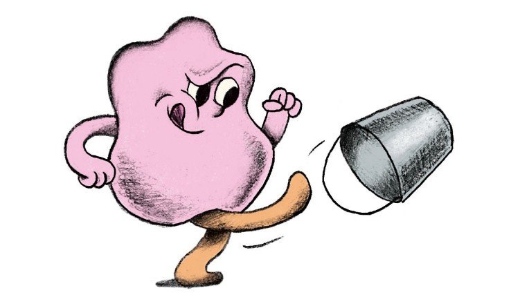
Illustration by Kinga Offert
The bucket-list idea is “a really cool concept”, says Petr Broz, an immunologist at the University of Lausanne in Switzerland, although he notes that “it’s hard to prove experimentally”.
Even once cell death begins, it isn’t always inevitable, says Ho Lam Tang, a cell biologist at Johns Hopkins University School of Medicine in Baltimore, Maryland. Tang and his sister, Ho Man Tang, a tumour biologist and postdoc in another laboratory at Hopkins, observed this while studying apoptosis at the Chinese University of Hong Kong in 2007. At the end of their experiments, they noticed that tiny, struggling cells remained in the culture dishes. Curious, one night they washed away the cell-death-inducing agent and incubated the shrunken cells in fresh medium. The next morning, to their surprise, many of the cells had recovered.
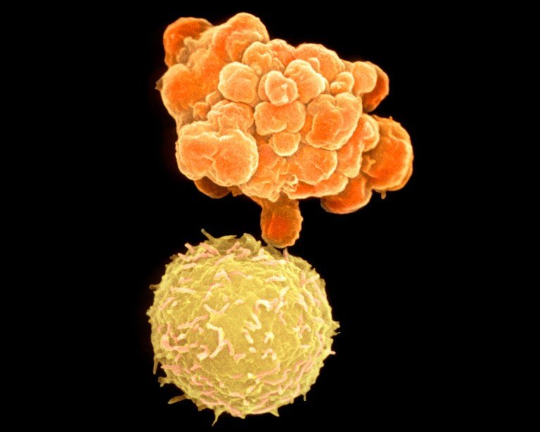
Two human white blood cells; one (top) is undergoing apoptosis, a programmed form of cell death.Credit: Dr Gopal Murti/Science Photo Library
The cells seemed to have activated various repair pathways and resurrected themselves, in a process the researchers called anastasis, Greek for ‘rising to life’. Their assertion countered the prevailing dogma that apoptosis, once initiated, was irreversible. The siblings submitted their study to 11 journals before a video of the reviving cells convinced an editor at Molecular Biology of the Cell to publish the work8, in 2012. Since then, the researchers have observed reversal of ferroptosis and necroptosis, and they anticipate that it will be possible after other forms of cell death.
A guide to cell death pathways
The occasional reversibility of cell death suggests that the point of no return is much later than scientists once thought. Both Broz and García-Sáez speculate that it comes once the borders of a cell’s mitochondria are irreparably disrupted, obliterating its metabolism. “At some point,” García-Sáez says, “when it’s dead, it’s dead.”
For more tech updates, stay tuned to our blog.