Mouse models The animal experimental procedures were conducted in strict compliance with the guidelines established by Weill Cornell Medicine, following the Guide for the Care and Use of Laboratory Animals, and the standards set by the Association for Assessment and Accreditation of Laboratory Animal Care International. The Research Animal Resource Center, Weill Cornell Medicine’s Institutional
Mouse models
The animal experimental procedures were conducted in strict compliance with the guidelines established by Weill Cornell Medicine, following the Guide for the Care and Use of Laboratory Animals, and the standards set by the Association for Assessment and Accreditation of Laboratory Animal Care International. The Research Animal Resource Center, Weill Cornell Medicine’s Institutional Animal Care and Use Committee and the Cornell Institutional Animal Care and Use Committee reviewed and approved all procedures for the mouse study under protocols 2011-0031 (principal investigator (PI): D.B.) and 2025-0008 (PI: D.B.). Mice were maintained under a 12-h light–dark cycle at 20–22 °C and 40–60% relative humidity in a specific pathogen-free facility with ad libitum access to food and water. Experiments involved both male and female mice and were sex-matched and age-matched (ranging from 8 to 16 weeks) unless otherwise specified. All experimental groups included both sexes. No sex-based differences or biases were observed. Arid1a (strain 027717), Cg1cre (strain 010611) and Pbrm1 (strain 031875) mice were sourced from The Jackson Laboratory. The CtcfHet (Ctcf+/-Cg1cre/+) mouse strain was a gift from the laboratory of G. Lauvau. The Arid1a strain was back-crossed to the C57BL/6J background for at least six generations before being used in experiments. In all experiments the Cg1cre/+ allele was used (heterozygous).
Sample sizes were determined on the basis of standards in the field and previous experience with high-throughput sequencing and flow cytometry experiments. ATAC–seq and RNA-seq experiments were performed with multiple biological replicates (n = 2–4 per condition). Littermates were randomly assigned to experimental groups where feasible. All genetic combinations (allelic genotypes) were derived from the same breeding pairs and analysed as littermates to control for genetic background and environmental variation, thereby minimizing potential bias. Experiments included 3–5 animals per group unless otherwise specified. For sequencing experiments (ATAC–seq and RNA-seq), samples were coded and assigned randomized identifiers before experiments and library preparation. Investigators handling and processing samples were blinded to the genotype or treatment group during these steps. For other assays (for example, proliferation or immunoblotting), blinding was not performed; however, objective quantification methods and standardized pipelines were applied uniformly across conditions to minimize bias.
Cell lines
Mammalian cell lines were maintained at 37 °C in a humidified environment comprising 5% carbon dioxide. Cell growth media were supplemented with 2 mmol l–1 GlutaMAX (35050061, Gibco), 10 mmol l–1 HEPES and 1% penicillin G and streptomycin, as well as fetal bovine serum (FBS; A5256801, Gibco), unless indicated otherwise, in the concentrations specified below. Routine examinations were performed in the laboratory to check for any potential mycoplasma contamination in the cell lines. The hES cell research was conducted under the oversight of the ESCRO committee and approved under protocol number 2022-05-001.
HEPG2, ID8 and WI-38 cell lines, as well as HEK293T for virus production, were propagated in Dulbecco’s modified Eagle medium (DMEM) with 10% FBS. K562, KG1 and RIVA (RI-1) cell lines were propagated in Iscove’s modified Dulbecco’s medium (IMDM) with 20% FBS. PC3 and SVHUC lines were cultured in Ham’s F-12K (Kaighn’s) medium with 20% FBS. SUM159 cells were cultured in Ham’s F12 with 10% FBS, insulin (5 μg ml–1) and hydrocortisone (1 μg ml–1). OCI-AML3 and OCI-AML2 cell lines were cultured in alpha-MEM supplemented with 20% FBS. SHSY5Y cells were cultured in DMEM, supplemented with 10% FBS, non-essential amino acids (1:100; 11140050, Gibco) and sodium pyruvate (1:100; 11360070, Gibco). Raji, Jurkat, SK-MEL28 and SK-MEL239 cell lines were cultured with Roswell Park Memorial Institute (RPMI) medium supplemented with 20% FBS. The hES MEL-1 cell line was cultured in essential 8 Flex Base medium and essential 8 Flex 50× supplement (A2858501, Thermo Fisher) with normocin (ant-nr-05, InvivoGen).
The S2 Drosophila melanogaster cell line was maintained at 24 °C in a non-humidified, non-CO2-adjusted atmosphere, cultured in Schneider’s Drosophila medium supplemented with 10% FBS.
All cell lines were obtained from established repositories or collaborators. Specific sources include Accegen (OCI-AML2), the American Type Culture Collection (HEK293T, HEPG2, Jurkat, K562, PC3, Raji, S2, SHSY5Y, SK-MEL28, SVHUC and WI-38), Cytion (SUM159) and DSMZ (RIVA and OCI-AML3). The hES cell line MEL-1 was obtained from G. Macdonald, SK-MEL239 and KG1 cell lines were obtained from the laboratory of D. Landau, and ID8 was obtained from the laboratory of N. B. Chetrit. Cell lines were authenticated by the providers or via STR profiling where available. Cell lines were routinely tested for mycoplasma contamination. The results of the testing were negative.
Drug treatments
Cells were treated with the indicated concentrations unless indicated otherwise in the figures: 5 μM BRD-K98645985 (MedchemExpress), 100 nM FHD-286 (Foghorn Therapeutics), 5 nM FHD609 (Foghorn Therapeutics), 5 nM FHT006710 (Foghorn Therapeutics)91 or 5 μM PBRM1-BD2-IN-8 (MedchemExpress). Control cells were treated with an equivalent volume of vehicle (DMSO). ATAC–seq and ChIP–seq experiments were performed after 24 h of treatment, unless specified otherwise.
Generation of knockout cell lines using CRISPR–Cas9
To generate the double (CTCF and ARID1A, and ARID2 and ARID1A) knockout models in lymphoma cell lines, we used RIVA (RI-1) lines, including the RIVA cell lines with the ARID1A gene reverted to the WT status, using a previously described method24. To generate a breast cancer model with ARID1A deletion, we used the WT SUM159 cell line. The cells were electroporated with a ribonucleoprotein complex, which contained the Alt-R recombinant S.p. HiFi Cas9 nuclease (IDT 1081061), Alt-R CRISPR-Cas9 tracrRNA (IDT 1072534) and Alt-R CRISPR–Cas9 crRNAs, with the crRNAs targeting the following genes: CTCF: CD.Cas9.BNYC8101.AA (5′-CATGCTCGGTTTACCCAAAG-3′) or CD.Cas9.JZFQ4398.AA (5′-ATGCCGAACCAATTCTCCA-3′); ARID2: Hs.Cas9.ARID2.1.AB: (5′-ATTTCATCCACCTCGAAAGC-3′); ARID1A: targeting the precise Q474* mutation region (5′-GGACTTTACTGGTTGTAATA-3′), as well as the WT DNA template (5′-*A*C*AAGGCCCCAGCGGGTATGGTCAACAGGGCCAGACTCCATATTACAACCAGCAAGTCCTCACCCTCAGCAGCAGCAGCCACCCTACTCCCAGCAA*C*C*A-3′) (asterisks indicate phosphorothioate nucleotides that were modified to ensure DNA stability); or SMARCA5: Hs.Cas9.SMARCA5.1.AB (5′-GTCAGAGACGTTTTATTACC-3′). Electroporation was carried out in accordance with the manufacturer’s protocol using a SF Cell Line 96-well Nucleofector kit (V4SC-2096) with the CM-137 program. At 48 h after electroporation, cells were sorted to collect the live population (DAPI-negative after DAPI stain). Given the lethality of CTCF deletion, the CTCF-deficient and ARID1A-deficient cells were monitored for viability with trypan blue exclusion assay, sorted 48 h after electroporation and immediately subjected to ATAC–seq, whereas the remaining cells were used for immunoblotting to verify the efficiency of deletion. In other cases, cells were single-cell sorted into 96-well plates to allow clonal colony formation and generation of stable cell lines. Deletion efficiency was confirmed by immunoblotting before performing further experiments.
For CRISPR–Cas9-mediated deletion of osa in S2 cells, we used Alt-R CRISPR-Cas9 crRNAs: CD.Cas9.SKCP0295.AB (5′-TGTTGACCAACACCCTGTGA-3′) and CD.Cas9.CMBJ6893.AD (5′-GACCAAGAGCAAGACCTGGA-3′). Electroporation was performed using a SF Cell Line 96-well Nucleofector kit (V4SC-2096) with the DS-137 program. The cells were subjected to ATAC–seq 48 h after electroporation, and deletion efficiency was confirmed by immunoblotting.
Generation of CTCF-overexpressing and ARID1A knockdown cell lines using lentiviral transduction
RIVA cell lines stably overexpressing CTCF were generated via lentiviral transduction using pHAGE-CTCF vector (Addgene, 116728). RIVA ARID1A knockdown cells were generated using lentiviruses previously generated in earlier work24. The following antisense guide sequences were used to target ARID1A: ARID1A-7170: TATGTTTACAGTATCAGGTGTA, ARID1A-6964: TTACCATTAAAAACAGTTATGA. Lentiviral particles were produced by transfecting HEK293T cells with the packaging plasmids psPax2 (Addgene, 12260) and psMD2.G (Addgene, 12259) together with the CTCF expression vector at a 4:3:1 ratio. Viral supernatants were collected at 48 and 72 h after transfection, pooled, filtered through a 0.45 μm membrane and concentrated using PEG-it according to the manufacturer’s instructions (LV825A-1, System Biosciences). The concentrated virus was resuspended in PBS supplemented with 25 μmol l–1 HEPES and used to infect RIVA cells. Following transduction, GFP-positive cells were isolated by sorting, and CTCF overexpression was validated by immunoblotting.
Generation of hES cells with an ARID1A degron
The endogenous hES cell ARID1A allele was tagged with dTAG by introducing a FKBPV-2×HA-P2A-NLS-eGFP cassette to the ARID1A carboxy terminus. The construct, including the FKBP12 degron flanked by homology arms 797 bp upstream and 800 bp downstream of the ARID1A C terminus, was ordered from Integrated DNA Technologies. The vector was introduced into MEL-1 hES cells together with the pX330 Cas9 vector and guide RNAs (TGTCCCACGGGCTGTCATGAC, TCATGACAGCCCGTGGGACAC) targeting the ARID1A stop codon using lipofection in mTeSR medium mixed with CloneR solution (1:10). Transfected cells were selected for 48 h with G418 (500 μg ml–1) using mTeSR:CloneR (1:10). After selection, cells were single-cell sorted based on the eGFP reporter fluorescence intensity, and only the high-eGFP population was used for the formation of single-clone colonies. Clonal lines were expanded and characterized by genotyping PCR, and selected homozygous clones were further validated by immunoblotting.
Isolation of MEFs and Cre-mediated gene deletion
MEFs were isolated from either male or female embryonic day 13.5–14.5 embryos obtained from pregnant mice carrying floxed alleles of the gene (or genes) of interest. MEFs were confirmed to be mycoplasma-free and cultured at 37 °C in a humidified incubator with 5% CO2 and low O2 (4%). The MEFs were maintained in DMEM (Corning, 10-013-CV) supplemented with 20% FBS (R&D Systems, S10250 or Gembio, 100–106), 2 mM GlutaMAX (Gibco, 35050061), 0.1 mM MEM non-essential amino acids (Gibco, 11140-050), 100 U ml–1 penicillin, 100 μg ml–1 streptomycin (Gibco, 15140163) and 55 μM β-mercaptoethanol (Gibco, 21985023). For genetic deletion, MEFs were subjected to Cre recombinase-mediated excision of the floxed allele. Cells at p.1 were infected with pMSCV-Cre-IRES-GFP retroviral vector to induce excision, followed by sorting to enrich GFP-positive cells. Control cells were processed similarly with an empty pMSCV-IRES-GFP vector infecting dividing cells, rather than GFP-negative cells to avoid selecting cells with different proliferation potential. Efficient gene deletion was confirmed by PCR genotyping.
Immunoblotting
For immunoblotting, cells were lysed in RIPA buffer (20 mM HEPES KOH pH 7.5, 50 mM β-glycerophosphate, 1 mM EDTA pH 8.0, 1 mM EGTA pH 8.0, 0.5 mM Na3VO4, 100 mM KCl, 10% glycerol and 1% Triton X-100, supplemented with complete protease inhibitor cocktail; Roche, 05056489001). The concentration of proteins in the supernatant was assessed using Bradford reagent (5000205, Bio-Rad) and absorbance acquisition. Lysates were subject to resolution through SDS–PAGE, then transferred to a PVDF membrane overnight. The membrane was subsequently blocked with 5% milk (Bio-Rad 1706404) and probed using primary antibodies in concentrations listed below. The membrane was then incubated with a corresponding peroxidase-conjugated secondary antibody (1:3,000 dilution Cell Signaling Technology (CST), 7074 and 7076), and detected with chemiluminescence substrate (34095, Thermo Scientific) on a ChemiDoc MP Imaging system (Bio-Rad). Densitometry values were subsequently obtained using Fiji software (National Institutes of Health). The following antibodies were used: ACTL6A (A301-391, Bethyl Laboratories; 1:3,500); ARID1A (HPA005456, Sigma Aldrich; 1:1,000); ARID2 (166117, Santa-Cruz; 1:200); BRD7 (15125, CST; 1:500); BRD9 (A303-781A, Bethyl Laboratories; 1:5,000); CTCF (3418, CST; 1:1,000); DPF2 (ab134942, Abcam); GAPDH (97166, CST; 1:2,000); Osa (AB_528420, DSHB); PBRM1 (A301-591A, Bethyl Laboratories; 1:2,000); SMARCA2 (sc-17828, Santa Cruz; 1:500); SMARCA4 (13-2002, EpiCypher; 1:5,000); SMARCB1 (91735, CST; 1:2,000); SMARCD1 (sc-135843, Santa-Cruz; 1:500); SMARCE1 (A300-810A, Bethyl Laboratories; 1:5,000); SMC3 (A300-060A, Bethyl Laboratories; 1:10,000); SMARCA5 (SNF2H) (A301-017A, Bethyl Laboratories; 1:2,000); human tubulin (T0198-25UL, Sigma Aldrich; 1:5,000); and D. melanogaster tubulin (AB_2732839, DSHB).
Protein IP
For FHD-286 treatment, cells were treated for 24 h before collection. Protein IP was performed in nuclear lysates. Nuclei were isolated from 200 × 106 cells following the ‘Nuclei Isolation for Single Cell Multiome ATAC+ Gene Expression Sequencing’ protocol (10x Genomics) with the following modifications: the cell membrane was lysed with 20 ml of the lysis buffer, followed by lysis inactivation with 30 ml of the wash buffer and then another wash in 30 ml of wash buffer. Isolated nuclei were then lysed in 4 ml lysis buffer (20 mM Tris, pH 7.5, 150 mM NaCl, 1 mM EDTA, 1 mM EGTA, 1% Triton X-100, 2.5 mM sodium pyrophosphate and 1 mM β-glycerophosphate) supplemented with protease and phosphatase inhibitors and incubated on ice for 30 min. Lysates were briefly sonicated (3 cycles of 8 s on, 30 s off; Diagenode Bioruptor Pico) and cleared by 10 min of centrifugation at 14,000g. The supernatant was incubated with an antibody (CTCF (3418, CST 4 µg), PBRM1 (A301-591A, Bethyl; 12 µg), BRD7 (A302-304A, Bethyl; 20 µg), IgG (13-0042, EpiCypher) or C15410206 (Diagenode), equal amount to the targeting antibody) overnight at 4 °C with rotation. For the experiment involving Benzonase (E1014, Millipore), 100 U of the enzyme and MgCl2 to a final concentration of 1.5 mM were added to the samples. After overnight incubation, protein A/G magnetic beads (26162, Invitrogen) were added and incubated for additional 4 h to capture target-interacting complexes. For immunoblot detection, beads were washed 3 times with the lysis buffer before elution in 2× Laemmli sample buffer and incubation in 95 °C for 5 min. For MS analyses, beads were washed 3 times with the lysis buffer, 3 times with cold PBS and frozen at −80 °C.
MS analysis
CTCF-interacting peptides, isolated from nuclear lysate by IP, were loaded on a 50 cm column (Thermo Fisher ES903) and separated by reverse-phase chromatography using a gradient from 1% to 30% buffer B over 130 min, to 90% buffer B over 5 min and held to 90% buffer B for 10 min with a flow rate of 300 nl min–1 using a Vanquish Neo system (Thermo Fisher). Buffer A was 0.1% formic acid (FA) in HPLC-grade water, buffer B was 0.1% FA in acetonitrile. The column temperature was maintained at 60 °C. MS analysis was conducted using an Orbitrap Fusion Lumos Tribrid mass spectrometer operating in data-independent acquisition mode with positive ionization. Full MS spectra were acquired in the m/z range of 380–985 at a resolution of 120,000, with standard automatic gain control (AGC) target settings and a maximum injection time of 50 ms. Targeted MS2 spectra were acquired in the m/z range of 145–1,450 using a precursor mass range of m/z 380–985, a quadrupole isolation window of 10 m/z, and 60 scan events. The MS2 scans were acquired at a resolution of 15,000 with an AGC target of 1 × 10⁵ and a maximum injection time of 40 ms. Fragmentation was performed using higher-energy collision-induced dissociation with a one-step collision energy of 30%. The electrospray voltage was maintained at a static setting, and the capillary temperature was set to 305 °C. No sheath or auxiliary gas flow was applied. IP PBRM1 samples were prepared for MS using S-Trap micro columns (ProtiFi) according to the manufacturer’s recommended protocol. Proteins were reduced using 20 mM tris((2-carboxyethyl) phosphine) for 30 min at 47 °C with gentle agitation and alkylated with 20 mM iodoacetamide for 30 min at room temperature in the dark. Then, samples were acidified with phosphoric acid at a final concentration of 1.2%. Samples were digested with 1 µg of sequencing-grade modified trypsin (Promega) for 3 h at 47 °C. Peptides were eluted sequentially with 50 mM triethylammonium bicarbonate, 0.2% FA and 0.2% FA in 50% acetonitrile. Eluted peptides were dried and resuspended in loading buffer reverse phase (0.1% FA in HPLC-grade water). The peptides were loaded on a 25 cm × 75 µm x 1.5 µm PepSep C18 HPLC column with a 10 μm CaptiveSpray emitter using a NanoElute 2 system (Bruker) with a linear gradient of 3–20% buffer B over 90 min followed by a step from 20–37% buffer B in 20 min at 150 nl min–1 and 37–95% buffer B in 10 min at 200 nl min–1 and held at 95% for 5 min. The gradient was then decreased to 5% buffer B for 5 min at 200 nl min–1. Buffer A was 0.1% FA in HPLC-grade water, buffer B was 0.1% FA in acetonitrile. The column temperature was maintained at 50 °C. The timsTOF Ultra mass spectrometer (Bruker) was operated in data-independent acquisition parallel accumulation, serial fragmentation mode, using variable-width ion mobility-m/z windows. There were no overlap windows in m/z, but ion mobility windows were acquired with varying overlaps. TIMS settings were 100 ms ramp and accumulation time (100% duty cycle) and ramp rate of 9.43 Hz; this resulted in 1.17 s of total cycle time, and mass spectra were recorded in the m/z range of 300–1,470 for the bulk samples, whereas 0.95 s of total cycle time and mass spectra were recorded in the m/z range of 400–1,000. Collision energy was applied in a linear fashion, with an ion mobility of 0.6–1.6 V s cm–2 and collision energy from 20 eV to 59 eV.
Proliferation assay
Cell proliferation was assessed using a CellTiter-Glo Luminescent Cell Viability assay (G7572, Promega) over a period of 5 days according to the manufacturer’s manual. Cells were seeded in 384-well plates with transparent bottom at a density of 4,000 cells per well for suspension cells (RIVA) and 500 and 1,500 cells for adherent cells (MEFs) in 50 μl of appropriate culture medium. Luciferase-containing lysis buffer was added to a portion of cells for luminescence measurement at the time of seeding and then every 24 h using a BioTek Synergy Neo plate reader (Agilent). Background luminescence from cell-free wells was subtracted from all readings. The relative luminescence intensity, which correlates to ATP levels and viable cell number, was normalized to 0 h values and expressed as the fold change over time. Three to six technical replicates were measured for each time point for each cell line.
Bulk RNA-seq
Expression levels of genes encoding CTCF and cohesin subunits and PBAF subunits were assessed by bulk RNA-seq. RNA was isolated from non-treated RIVA cells using a RNeasy Mini kit (74104, Qiagen) according to the manufacturer’s manual. RNA samples were submitted to The Weill Cornell Genomics Resources Core Facility for RNA-seq library preparation, followed by sequencing with a NovaSeq 6000 System.
Bulk ATAC–seq
Experiments were performed in biological triplicate for each condition. When three independent clones were not available, three parallel cultures of the same cell line were maintained separately for at least 7 days before the experiment. This approach ensured that replicates were derived from independently propagated cell populations rather than representing technical replicates. Cells were passaged 24 h before the procedure to ensure logarithmic growth at the time of the experiment. For ATAC–seq following pharmacological treatment, cultures were passaged again after 24 h and split in two, with one fraction treated with a drug, whereas the other received an equivalent volume of DMSO as a control. In the case of ATAC–seq after treatment, after 24 h, the culture was split into two, and one part received a drug and the other DMSO, followed by 24 h of treatment before the experiment. Cells were incubated for 24 h before ATAC–seq analyses. For ATAC–seq of mouse primary cells, freshly sorted cells were isolated from mouse spleens and processed immediately. The procedure followed the Omni-ATAC protocol92. In brief, 75,000 live cells were rinsed in cold PBS and lysed in 50 μl cold lysis buffer (10 mM Tris-HCl, pH 7.5, 10 mM NaCl, 3 mM MgCl2, 0.1% NP-40, 0.1% Tween-20 and 0.01% Digitonin) for 5 min on ice. Next, 1 ml wash buffer (10 mM Tris-HCl, pH 7.5, 10 mM NaCl, 3 mM MgCl2 and 0.1% Tween-20) was added, and the samples were centrifuged. The pelleted nuclei were then resuspended in a transposition reaction mix that was prepared using Illumina Tagment DNA TDE1 Enzyme and Buffer kits (Illumina 20034197) and incubated at 37 °C for 30 min on a thermomixer set at 1,000 rpm. Tagged DNA fragments were purified using a Clean & Concentrator-5 kit (Zymo D4014) and then subjected to PCR amplification to generate sequencing libraries. PCR amplicons were purified using a Clean & Concentrator-5 kit (Zymo D4014). The size distribution and quality of the library were evaluated using a High Sensitivity DNA kit (Agilent 5067-4626) and run on an Agilent 2100 Bioanalyzer. The libraries were sequenced on Illumina NovaSeq 6000 and NovaSeq X Plus platforms using a PE50 SP kit.
ChIP–seq
ChIP–seq was carried out as previously described24 with slight modifications. Changes to the protocol were as follows: (1) hES cells were double-fixed with 2 mM DSG (Sigma 80424-5MG-F) for 50 min, followed by 1% formaldehyde at room temperature for 10 min; (2) double-fixed mES cells were added to each hES cell sample in equal amount as a spike-in control before cell lysis; (3) the number of chromatin sonication cycles was adjusted per experiment to maintain consistent shearing pattern, ranging from 10 to 26 cycles of 30 s of sonication in Diagenode Bioruptor Pico, with 30 s of breaks in between cycles; and (4) DNA was purified using chloroform–phenol extraction. Immunoprecipitated DNA and input DNA were subject to library preparation (NEBNext Ultra II DNA Library Prep kit, Illumina). The following antibodies were used: ARID1A (ab182560, Abcam), BRD7 (A302304AT, Bethyl Laboratories), CTCF (91285, Active Motif), DPF2 (ab134942, Abcam), HA (3724, CST), H3K4me1 (ab8895, Abcam), PBRM1 (A301-591A, Bethyl Laboratories) and SMC3 (5696, CST, for RIVA and SUM159 cells, ab9263, Abcam, for hES cells). Libraries for next-generation sequencing were prepared using a NEBNext Ultra II DNA Library Prep kit for Illumina (New England Biolabs, E7645L) with dual indexes (E6440, NEB). Purification of DNA was carried out using AMPure XP beads (NC9933872, Thermo Fisher Scientific). The concentration was measured using a Qubit Fluorometer (Thermo Fisher) with a dsDNA High Sensitivity kit (Q32851), and the size of DNA fragments was assessed with a High Sensitivity DNA kit (Agilent 5067-4626) on an Agilent 2100 Bioanalyzer. The libraries were sequenced on Illumina NovaSeq 6000 and NovaSeq X Plus platforms using a PE50 SP kit with minimum 20 million reads per sample.
Micro-C
Micro-C libraries were prepared following a published protocol51,93 with minor modifications. First, the dinucleosome band was size-selected using an E-gel EX 2% agarose (A42135, Invitrogen) after DNA purification. Second, the end-repair step using the Lucigen End-It DNA End-Repair kit (now discontinued) was omitted. Adjustments were applied to two experimental setups: (1) RIVA WT and ARID1AHet and (2) RIVA WT, ARID1AHet and ARID1AHetARID2Hom, as detailed below.
In brief, 30 × 106 RIVA cells were crosslinked with 1% formaldehyde for 10 min at room temperature, followed by quenching with 750 mM Tris-HCl (pH 7.4). Fixed cells were washed with PBS, further crosslinked with 3 mM DSG for 45 min, quenched with 750 mM Tris-HCl and washed again. The doubly crosslinked cells were counted and pelleted in 1 × 10⁶ aliquots and flash-frozen and stored at −80 °C until proceeding further.
Micrococcal nuclease (MNase, NEB M0247S) digestion was determined by titration to achieve an 80:20 to 90:10 mononucleosome to dinucleosome ratio. Cells were resuspended in MB1 buffer (10 mM Tris-HCl pH 7.5, 50 mM NaCl, 5 mM MgCl2, 1 mM CaCl2 and 0.2% NP-40) supplemented with protease inhibitor cocktail and 1× PMSF and digested with the MNase amount pre-optimized for each batch (3.25 μl NEB MNase M0247S for experiment (1) and 0.2 μl for experiment (2)) at 37 °C for 10 min. Digestion was stopped by adding 0.8 mM EGTA and heating to 65 °C for 10 min. The nuclei were then washed with MB2 buffer (10 mM Tris-HCl pH 7.5, 50 mM NaCl and 10 mM MgCl2).
The DNA ends were repaired with T4 PNK (M0201, NEB) and filled in with biotinylated nucleotides (biotin-14-dATP;NU-835-BIO14-L and biotin-11-dCTP;NU-809-BIOX-L, Jena Biosciences) using Klenow (large fragment) DNA polymerase (M0210, NEB). Proximity ligation was performed at 25 °C for 2.5 h for experiment (1) and overnight for experiment (2) using T4 DNA ligase (M0202S, NEB), followed by exonuclease III (M0206, NEB) treatment to remove unligated biotinylated ends. Crosslink reversal, RNase A and proteinase K treatment were performed in a single step at 65 °C overnight for experiment (1) and for 9 h for experiment (2), followed by DNA purification using phenol–chloroform–isoamyl alcohol extraction. The aqueous phase was further processed using a Zymo Clean & Concentrate 5 kit (D4014, Zymo) and eluted in 20 µl TE buffer.
Purified DNA was separated on an E-gel EX 2% agarose (A42135, Invitrogen), and the di-nucleosome band (around 250–400 bp) was excised and extracted using a Zymo Gel DNA Recovery kit (D4001). For library preparation, the DNA was bound to MyOne Streptavidin C1 beads (65001, Invitrogen), washed with TBW (10 mM Tris-HCl pH 7.5, 1 M NaCl, 0.5 mM EDTA and 0.1% (v/v) Tween-20) and processed using a NEB Ultra II DNA Library Prep kit (E7645L). Libraries were amplified with ten PCR cycles, quantified via BioAnalyzer and Tapestation, then sequenced on a NovaSeq X Plus platform using a PE150 SP kit to achieve at least 420 million reads per sample.
Bone marrow transplantation
Bone marrow cells were collected from the femur and tibia of donor mice aged between 8 and 12 weeks, followed by treatment with a red blood cell lysis solution (Qiagen 158904). Lethally irradiated C57BL/6J host mice, who received two doses of 450 RAD via a Rad Source Technologies RS 2000 Biological Research X-ray Irradiator, were then injected with 1 million of these cells through the retro-orbital sinus. These transplanted mice were used for experiments 8–16 weeks after transplantation. Excluding the mice euthanized at predetermined intervals, all mice participating in lymphomagenesis research were observed until they exhibited one or more conditions warranting euthanasia. These conditions included significant lethargy and body weight loss as well as discernible splenomegaly. This procedure aligns with the guidelines of our animal protocol approved by the Weill Cornell Medicine Institutional Animal Care and Use Committee (protocols 2011-0031 and 2025-0008, PI: Barisic).
Immunization
To induce germinal centre (GC) formation in the spleens, mice were immunized with an intraperitoneal injection of 500 μl of 2% sheep red blood cells resuspended in sterile 1× DPBS, prepared from a solution of sheep blood in Alsever’s (Cocalico Biologicals 20-1334A). Alternatively, mice were immunized via intraperitoneal injection with 80 μg NP(30-32)-KLH (Biosearch Technologies N-5060) adsorbed in alum adjuvant (ThermoFisher 77161) at a 1:1 ratio of alum to immunogen before injection.
Flow cytometry of GC B cells
Spleens were homogenized, and splenocytes were filtered through a 40 μm cell strainer, followed by enrichment of mononuclear cells using density gradient centrifugation with Ficoll–Paque Premium (Cytiva 17-5442-02). The resulting single-cell suspensions were resuspended in PBE (PBS with EDTA) containing 0.5 μg ml–1 rat anti-mouse CD16/CD32 (clone 2.4G2, BD) and incubated for at least 5 min on ice to block Fc receptors. Without washing, cells were stained for 20 min on ice with a mix of fluorochrome-conjugated anti-mouse antibodies, either diluted in PBE or in a 50:50 mix of Brilliant stain buffer (BD 566349) and PBS with 0.5% BSA (without EDTA), when the mix included ≥2 BV-conjugated or BUV-conjugated antibodies, to prevent staining artefacts caused by dye interactions. The cells were washed twice before analysis. DAPI (ThermoFisher D1306) was added immediately before acquisition at a final concentration of 0.5 μg ml–1 to exclude dead cells. Data were acquired on a BD Symphony A3 and analysed using FlowJo software. The following antibodies and dilutions were used: B220-APC (clone RA3-6B2, BD 553092; 1:100); B220-BV786 (clone RA3-6B2, BD 563894; 1:300); CD138-BUV737 (clone 281-2, BD 564430 and BD 624230; 1:500), CCR6-PerCP-Cy5.5 (clone 29-2L17, BioLegend 129809; 1:300); CD11b-APC (clone M1/70, BioLegend 101212; 1:400); CD21-APC (clone 7E9, BioLegend 123412; 1:500); CD21-PECy7 (clone 7E9, BioLegend 123420; 1:500); CD4-BUV563 (clone GK1.5, BD 612923; 1:500); CD38-BUV395 (clone 90, BD 740245; 1:500), CD38-BUV563 (clone 90/CD38, BD 741271; 1:400); CD80-BV421 (clone 16-10A1, BioLegend 104725; 1:300); CD86-BV605 (clone GL-1, BioLegend 105037; 1:200); CXCR4-biotin (BD 551968; 1:200); CXCR4-PE (clone 2B11,Invitrogen 12-9991-82; 1:100); ephrin-B1-biotin (R&D Systems, BAF473; 1:100); FAS-BUV805 (clone Jo2, BD 741968; 1:100); FAS-PE-Cy7 (clone Jo2, BD 557653; 1:500); GL7-FITC (BD 553666; 1:500); GL7-PerCPCy5.5 (clone GL7, BioLegend 144610; 1:500); IgD-BV510 (clone 11-26c.2a, BD 563110; 1:500); IgD-PerCP (clone 11-26c.2a, BioLegend 405709; 1:500); IgG1-BV650 (clone A85-1, BD 740478; 1:500); IgM-BUV661 (clone II/41, BD 750660; 1:300); IgM-BUV805 (clone II/41, BD 749307; 1:500); kappa-APC-Cy7 (1:200), lambda-BV421 (1:400); and PD-L2-BUV395 (clone TY25, BD 565102; 1:75). The following secondary antibodies were used: SA-APC-Cy7 (BioLegend 405208; 1:100) and SA-BV605 (BioLegend 405229; 1:200). NP-PE (N-5070-1) was diluted to 5 µg ml–1.
Multiome single cell RNA-seq and ATAC–seq
Single-cell RNA-seq and single-cell ATAC–seq libraries were prepared using 10x Genomics Single Cell Multiome assay kits (10x Genomics; 1000230, 1000283, 1000494, 1000215 and 1000212) following the manufacturer’s guidelines. In brief, GC cells from splenocytes were sorted using antibody cocktails described above (Flow Cytometry), rinsed with 0.04% BSA PBS, and 100,000 cells underwent the manufacturer’s nucleus preparation procedure and a combination of regular and low-input protocol was used. About 20,000 nuclei were placed into a 10x Chromium X instrument. The remainder of the library preparation steps were conducted in accordance with the manufacturer’s protocol. Libraries were evaluated using an Agilent Bioanalyzer, quantified using a KAPA Library Quantification kit and sequenced on an Illumina Novaseq platform.
Density sedimentation analysis by glycerol gradient ultracentrifugation
In brief, 180 × 106 cells were subject to nucleus isolation by lysis in buffer containing 25 mM HEPES (pH 7.6), 25 mM KCl, 0.05 mM EDTA, 10% glycerol, 5 mM MgCl2, 0.1% NP-40, supplemented with 1 mM DTT, protease inhibitors and 1 mM PMSF on ice, following a 15-min centrifugation at 1,000g and 4 °C. The nuclei were resuspended in a buffer containing 10 mM HEPES (pH 7.6), 3 mM MgCl2, 100 mM KCl, 0.1 mM EDTA, 10% glycerol supplemented with 1 mM DTT and protease inhibitor, and lysed by the addition of ammonium sulfate to final concentration of 0.3 M. Proteins are separated from chromatin through ultracentrifugation at 4 °C, 150,000g for 30 min and precipitated in 0.3 g ml–1 ammonium sulfate for 20 min on ice. After another ultracentrifugation at 4 °C, 100,000g for 20 min, the nuclear lysate was resuspended in HEM buffer containing 25 mM HEPES (pH 7.9), 0.1 mM EDTA, 12.5 mM MgCl2 and 100 mM KCl, supplemented with 1 mM DTT and 1 mM PMSF. The protein concentration was assessed using a Bradford assay (5000205, Bio-Rad). Samples were diluted to 800 μg of proteins in 300 μl HEM buffer. The lysates were then carefully overlaid onto 7 ml of 10–30% glycerol gradient (diluted with HEM buffer), prepared in a 14 × 89 mm polyallomer centrifuge tube (331372, Beckman). Tubes were placed in a SW-40 swing bucket rotor and centrifuged at 4 °C for 16 h at 40,000 rpm in Beckman Optima XPN-100 ultracentrifuge. For SUM159, 21 fractions of 0.4 ml were collected. For RIVA, 6 fractions of 0.5 ml were collected, followed by 29 fractions of 0.2 ml. The proteins were precipitated with resin, 1/10 volume of each sample. The concentrated protein fractions were subsequently used in immunoblot analyses.
Computational analyses
Bulk ATAC–seq analysis
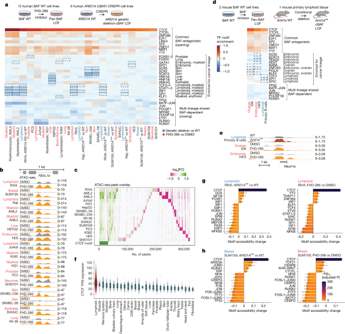
Raw sequencing reads were assessed using FastQC. Sequence adapters were removed with Trim Galore!, and the cleaned reads were aligned to either the mm10, hg38 or dm6 genome reference using BWA. Duplicate reads were identified with Picard94. Post-alignment, mitochondrial reads, duplicates, reads from blacklisted regions, secondary alignments, multimapping reads, reads with insert sizes greater than 2 kb, incorrectly oriented paired reads or orphan reads were removed. Peak calling was performed with MACS2 using a shift of −75 bp and an extension of 150 bp95. ATAC–seq quality control was done by evaluating fragment length distribution, FRIP scores, transcription start site (TSS) enrichment and high-quality autosomal reads with Picard, ataqv and MultiQC96,97. Consensus peaks were determined by overlapping regions covered in at least two samples. FeatureCounts was used to quantify reads in the peaks98. Data normalization for visualizations and unsupervised clustering was done using variance stabilizing transformation (vst)99. Differential peak accessibility was analysed using DESeq2 with a negative binomial model, filtering for an absolute fold change of 1.5 (log2[fold change] of 0.58) and an FDR-adjusted P value of 0.01. Peaks were annotated by identifying the most significantly changing peak within 5 kb of the TSS of each gene and assigning it to the promoter or potential enhancers of the gene, as defined by GREAT100. Genes were ranked by log2 fold change or the Wald statistic and analysed using GSEA with the MSigDB database and curated gene signatures from RNA-seq and literature101,102, filtering for adjusted P values less than 0.05. Transcription factor regulatory potential was evaluated by comparing mammalian motifs in JASPAR to sequences of accessible peaks103,104. A multivariate model was used to assess the relationship between log2 fold changes in peak accessibility and motif presence, adjusting for GC content, that is, log2[fold change = β0 + β1 × x1 + β2 × x2 + … + βN × GC, where xi is a Boolean variable for the presence of a TF motif in a peak as a binary variable and GC for the GC-content of the peak. Effect sizes and P values were derived using ordinary least squares regression with robust standard errors. Furthermore, TF-binding sites, binding scores and differential binding were analysed using TOBIAS footprinting105. Pseudomedians and 95% CI were calculated using the wilcox.test() function in R (v.4.3.1). Read density heatmaps were generated from normalized bigwig files using deepTools (v.3.5.5). Enrichment of CTCF knockout rescued and not rescued genes was performed using the hypergeometric distribution test and a curated list of CTCF-related signatures. The diversity of ATAC–seq response following FHD-286 treatment was assessed using interquartile range (IQR) of the DESeq2 log2 fold change at opening or closing (mean absolute fold change > 1.1 across all cell lines) peaks. CTCF motif overlap was determined using predicted CTCF motif sites from the JASPAR motif set, accessed through AnnotationHub record AH104727.
ChIP–seq analysis
Raw read quality control and adapter trimming were conducted using FastQC and TrimGalore!, followed by alignment of the reads to the hg38 genome reference with BWA106,107,108. Duplicate reads were identified using Picard94. After alignment, duplicate reads, reads in blacklisted regions, secondary alignments, multimapping reads, reads with an insert size over 2 kb, misoriented pairs or orphan reads were removed. Peaks were called for each IP sample compared with control samples using MACS2 (ref. 95). Consensus peaks were called as taking union of all the peaks. Peak calls were filtered to retain peaks present in at least two or three replicates, depending on the total number of samples in the experiment, with the required number not exceeding the number of replicates per condition minus one. Reads in peaks were quantified using FeatureCounts, and differential binding and annotation steps were performed similar to the ATAC–seq methods described above using DESeq2. In datasets with large copy-number differences between groups, CNVkit109 segment relative copy numbers were assigned to peaks, combined with DESeq2 library-size factors, and normalized using the DESeq2 normalizationFactors() function. Targets that had ChIP–seq experiments across multiple batches in the same conditions had their replicates combined when appropriate. Batch correction was performed using raw peak counts with a batch covariate in the negative binomial model of DESeq2, and plotting was performed using batch correction on the variance-stabilizing-transformed values using limma or on counts using ComBat-seq. ChIP–seq coverage was calculated using the bamCoverage function of the deeptools framework (v.3.5.5), using RPGC normalization where indicated and 10 bp bins. log2 fold change bigwigs were determined using the bigwigCompare function with pseudocount=1. Read density heat maps were prepared using the computeMatrix and plotHeatmap functions. PCA for all ChIP datasets generated in this study are shown in Supplementary Fig. 4b.
Multiome analysis
Multiome fastq files were aligned and processed using CellRanger Arc (v.2.0.0; 10x Genomics). The resulting outputs were further processed with Signac (v.1.5.0)110. Peaks were called using MACS2 with default settings95. A combined peak set was created from all samples, with peaks larger than 10 kb or smaller than 20 bases being filtered out. To correct for batch effects between experiments, Harmony (v.0.1.0) was applied on PCA (RNA) and LSI (ATAC) data111. Harmony-corrected values were used with Signac’s FindMultiModalNeighbors to generate multimodal nearest neighbours, which were used for UMAP visualization. Cell-type labels were assigned using RNA expression and the Seurat TransferLabel function with reference datasets70. Gene signature module scores were calculated using the AddModuleScore function with a control value of 5. Slingshot (v.2.8.0) was used to generate pseudotime, based on the first and second Harmony-corrected PCA of expression, starting from Centroblasts. The density and difference in density of cells for WT and ARID1AHet cells along pseudotime were calculated, with significance assessed using the Wilcoxon rank-sum test. ChromVAR scores for the JASPAR2020 core vertebrate motif list were calculated using Signac’s AddMotif and RunChromVAR functions on the filtered, combined peak set. Gene signature or ChromVAR accessibility of specific motifs were plotted against pseudotime, divided into ten deciles. Expression or accessibility for each decile was compared between WT and the indicated genotype using Wilcoxon rank-sum tests, and the differences in splines were plotted and coloured according to decile significance. Signac LinkPeaks was used to identify putative promoter–enhancer loops using the default settings, including a maximum distance of 500 kb for all genes. The multiome processing code can be accessed at GitHub (https://github.com/crchin/multiome_process).
Bulk RNA-seq analysis
Raw sequencing reads were processed using the nf-core/rnaseq pipeline112 (v.3.12.0) with default parameters. In the pipeline, Salmon (v.1.10.1) was used in quasi-mapping mode for transcript-level quantification against the GRCh38 human reference transcriptome (GENCODE v.33 annotation). Transcript abundances were imported into R and summarized to gene-level counts using the tximport package. Differential expression analysis was performed using DESeq2 (v.1.38.3). Genes encoding PBAF-specific subunits, as well as CTCF and cohesin subunits, were selected for targeted analysis. Count data were normalized using variance stabilizing transformation for plotting.
Micro-C analysis
Paired-end reads, obtained in biological duplicates for WT and ARID1AHet cells, were converted to valid pairs files and cool files following Dovetail’s Micro-C analysis protocol. The paired-end reads were aligned to the human genome (hg38) using BWA-MEM (v.0.7.18) with the following parameters: −5SP -T0. High-quality, valid pairs were retained using the parse module from the pairtools (v.0.3.0) implementing the following parameters: –min-mapq 40 –walks-policy 5unique –max-inter-align-gap 30. After the resulting pairsam file was sorted using pairtools sort, PCR duplicates were removed using the dedup module from pairtools with the following parameters: -mark-dups -output-stats. The pairtools split module was then used to generate a .bam and .pairs file. This process was performed individually on biological replicates and pooled replicates. Using the pairs file, two contact matrix file formats were generated: .cool and .hic. The .cool files were generated using cooler cload pairix (v.0.9.3) and KR-normalized using cooler zoomify –balance at 1 kb, 5 kb, 10 kb, 20 kb and 100 kb resolutions. The .hic files were generated using juicer tools (v.1.22.01) and KR-normalized at 10 kb and 20 kb resolutions. To ensure robust cross-condition comparisons and to remove systematic biases arising from sequencing depth, coverage heterogeneity and distance-dependence decay, we also jointly normalized replicate contact pairs for each genotype binned at 10 kb resolution using the fast loess procedure implemented in MultiHiCcompare (v.1.24.0). These fast-loess-normalized matrices were converted to .cool files. TADs were called using HiTAD (v.0.4.5) on 20 kb cool matrices and filtered to retain levels 0 and 1. Differential TAD domains were called using DiffDomain (v.1.0.0) on filtered HiTAD output. Insulation scores were obtained using the hicFindTADs tool from HiCExplorer (v.3.7.2) that were then mapped to boundaries from HiTAD TADs. Inter-TAD and intra-TAD connectivity across TAD boundaries was calculated using HiTAD output with the HiCExplorer hicInterIntraTAD function. Aggregate TAD plots were generated with the ATA function from the R package GENOVA (v.1.0.1) using 10 kb resolution loess-normalized matrices. GENOVA was also used to visualize contact matrices and differentials between WT and ARID1AHet data and to calculate relative contact probabilities. Loops were called using Mustache (v.1.0.1) on 5 kb KR-normalized .hic matrices per replicate. Pooled loops were obtained by the union of replicate loops. Loops were filtered using a P value threshold of 0.1 and a sparsity threshold of <0.6. To determine coordination of histone marks and gene expression in differential TAD domains, ChIP–seq mark summed densities were mapped to TAD domain regions, whereas gene expression was obtained from genes for which promoters overlapped with TAD domains. The coordination score was defined as previously described54. TAD boundary pile-ups were calculated using the union of TAD boundaries across the genotypes, and comparing the pile-ups to randomized shifted control regions using the shift option of coolpup.py.
Analysis of IP–MS data
Raw (.d) files were searched using the FragPipe computational platform (v.20.0) with the DIA-NN search engine (v.1.8.2_beta_8) against the UniProt Homo sapiens (UP000005640) database (reviewed sequences and isoforms; downloaded on 7 November 2023) using the BRUKER Human trypsin TIMScore (v.2) spectral library with a 1% FDR and default settings. A contaminant FASTA from the L. Hao Laboratory113 was included. The spectral library contains 13,114 protein groups and 573,437 precursor ions. Intensity data were log2-transformed and filtered to contain entries with at least two unique peptides and a minimum of 33.3% valid values in one group for comparisons. Missing values were imputed by random numbers drawn from a normal distribution, shifted down by 1.8 s.d. and with a width of 0.3 of each sample, using Perseus (v.2.0.11.0). Statistical analysis was performed using the R package Limma (v.3.60.3), applying an adjusted P value threshold (FDR-adjusted P < 0.05) to control for the FDR. Protein enrichment was determined relative to IgG control IPs.
Analyses of gene expression datasets
Gene expression data for normal tissue were retrieved from the Human Protein Atlas (HPA) on 26 July 2024. RNA expression was extracted from the single-cell RNA expression dataset (rna_single_cell_type.tsv) and grouped according to the cell-type classification from the HPA (rna_single_cell_type_cell_types.tsv), with groups containing fewer than three representative cell types excluded. Expression data were visualized by sorting cell-type groups based on median normalized TPM (nTPM) values. Gene expression data for human cancer cell lines was obtained from the DepMap Public 24Q4 dataset (accessed 26 July 2024). Expression values were log-transformed (log2[TPM + 1]) and merged with model metadata using ‘Cell Line Name’ as the common identifier. Lineages with fewer than three cell lines were removed, and median expression values were used to order cancer types. Gene expression data from human cancer cell lines were also retrieved from the HPA. Cell line information was extracted from the cell line dataset (rna_cell_lines.tsv), which was filtered to include only malignant cell lines. RNA expression data were extracted from the RNA gene expression dataset (rna_celline.tsv) and merged with the cell line dataset through the ‘Cell line’ identifier. Last, only rows corresponding to the gene of interest (CTCF) were retained. Cancer types with fewer than three representative cell lines were excluded, and nTPM values were summarized by median expression per disease type. All analyses were performed in Python (v.3.10) using pandas, seaborn and matplotlib.
For the DepMap dataset, the following violin sample sizes were used: lymphoid, n = 166; muscle, n = 5; nervous system, n = 40; myeloid, n = 62; bone, n = 43; bladder and urinary tract, n = 37; lung, n = 204; oesophagus and stomach, n = 73; uterus, n = 40; cervix, n = 20; CNS and brain, n = 97; bowel, n = 80; breast, n = 67; ampulla of Vater, n = 4; soft tissue, n = 56; liver, n = 27; pancreas, n = 54; prostate, n = 12; pleura, n = 22; skin, n = 95; ovary and fallopian tube, n = 67; kidney, n = 46; thyroid, n = 16; biliary tract, n = 42; testis, n = 6; head and neck, n = 65; eye, n = 19; and fibroblast, n = 39.
For the HPA datasets, sample sizes of normal cells were as follows: blood and immune cells, n = 12; squamous epithelial cells, n = 4; trophoblast cells, n = 3; muscle cells, n = 3; mesenchymal cells, n = 4; glandular epithelial cells, n = 18; specialized epithelial cells, n = 11; neuronal cells, n = 6; glial cells, n = 6; endocrine cells, n = 3; and germ cells, n = 5.
For malignant cells, the following sample sizes were used: lymphoma, n = 76; leukaemia, n = 93; neuroblastoma, n = 17; myeloma, n = 34; cervical cancer, n = 8; bone cancer, n = 21; bladder cancer, n = 26; gastric cancer, n = 42; rhabdoid, n = 14; colorectal cancer, n = 63; breast cancer, n = 62; pancreatic cancer, n = 46; sarcoma, n = 15; lung cancer, n = 232; oesophageal cancer, n = 27; prostate cancer, n = 8; liver cancer, n = 24; bile duct cancer, n = 7; ovarian cancer, n = 59; brain cancer, n = 80; uterine cancer, n = 29; skin cancer, n = 62; head and neck cancer, n = 38; thyroid cancer, n = 11; and kidney cancer, n = 35.
Multimark chromatin segmentation and clustering analysis
The chromatin segmentation, quantification and clustering steps were completed as previously described24. In brief, a common peak set was generated from the input marks (ATAC–seq, CTCF ChIP–seq and PBRM1 ChIP–seq). Peaks were extended to a minimum size of 500 bp and a graph was generated in which each vertex is a peak and two vertices are connected only when the two peaks overlap by >50% of their size in both directions. The graph was split into connected components, and components were collapsed into regions by taking the maximal extent of the peaks in that component. Any unconnected peaks were added to this set by their original coordinates. The start and end coordinates of all the regions were concatenated and taken as a set of boundary coordinates, and the genome was segmented into disjoint regions between these boundaries to make up the candidate segmentation set. The original peaks across all the marks and replicates were overlapped with this candidate set. Any region with fewer than two overlaps were discarded. Any adjacent region with similar peak presence or absence values (as a Hamming distance of binary values less than 2) were combined into one. After the peaks were segmented into these regions, subread featureCounts98 was used to quantify the reads of every mark across all the datasets. Normalized log-FPKM scores were calculated for each file, and the replicates were collapsed in a dataset by taking the median per region, and the log2 fold change of the differential (ARID1AHet versus WT) was calculated by taking the log-FPKM difference between the conditions. The normalized score for the WT conditions and the log2 fold change scores were clustered using k-means clustering using the input feature set used during peak segmentation, and the rest were used as independent read-outs. Gene expression was associated to the regions by taking the nearest TSS to each region and taking the expression difference. CTCF motifs were annotated using the CTCF R package and the EMBL M1 and M2 motifs114,115. For Supplementary Fig. 11, the motif enrichment was calculated taking the peaks in each cluster and assigning them a dependent variable of 1, and any other peak outside of that region as 0, and running the multivariate motif enrichment as described in the ATAC–seq section. The TAD insulation scores were calculated by taking the insulation scores around 100 kb of each region from Micro-C results with a 10 kb bin size, and shifting each profile vertically so that the maximal insulation score of each peak is shifted to zero. Therefore the insulation score difference between ARID1AHet and WT data would be calculated as the delta of the trough at the centre of the region (if any) compared with the background connectivity for each condition, and the differences in the extents of the dips can be calculated between ARID1AHet and WT.
Survival data analysis
Overall survival was calculated and graphically represented using cBioPortal (www.cbioportal.org) (PMID: 22588877). Studies included in the analysis are part of TCGA study (PMID: 24071849). Kaplan–Meier plots were cut once only one patient was left in one of the groups. Significance was determined by performing log-rank tests.
Quantification and statistical analyses
Statistical parameters such as the number of replicates (n), precision measures (mean or median ± s.d.) and significance are provided in the figures and figure legends. Results were considered significant if P < 0.05 or adjusted P < 0.05 according to the specified test. Statistical analyses were conducted using R (v.4.1.0) and Prism GraphPad (v.10.0). Details of the statistical analysis for sequencing data are outlined in the Methods and figure legends.
Inclusion and ethics statement
All animal studies were approved by the Institutional Animal Care and Use Committee of Weill Cornell Medicine under protocols 2011-0031 (PI: D.B.) and 2025-0008 (PI: D.B.), and conducted in compliance with institutional guidelines and the Guide for the Care and Use of Laboratory Animals. Both male and female mice were used in all experiments, with age-matched and sex-matched cohorts, and no sex-based differences were observed. All hES cell research was approved by the Weill Cornell ESCRO committee (protocol 2022-05-001). No human participants or identifiable human data were used.
Biological materials
All commercially available and previously published materials used in this study are referenced in the text. Unique biological materials generated in this study, including ARID1A-mutated lymphoma cell lines and Arid1aflox/floxCtcfflox/floxCg1Cre double conditional deletion mice, are available from the corresponding authors upon reasonable request and may be subject to a Material Transfer Agreement.
Reporting summary
Further information on research design is available in the Nature Portfolio Reporting Summary linked to this article.
Check back often for more exciting news!