tRNA in vitro production Oligonucleotides containing tRNA under a T7 promoter and followed by an HDV ribozyme61 were synthesized by IDT as high-fidelity DNA microchip oligo pools from 254–275 nucleotides in length, and dsDNA was amplified in a single PCR reaction for each pool. These tRNA pools included 48 unique E. coli isoacceptor tRNAs with
tRNA in vitro production
Oligonucleotides containing tRNA under a T7 promoter and followed by an HDV ribozyme61 were synthesized by IDT as high-fidelity DNA microchip oligo pools from 254–275 nucleotides in length, and dsDNA was amplified in a single PCR reaction for each pool. These tRNA pools included 48 unique E. coli isoacceptor tRNAs with all combinations of base 75, all combinations of bases 74 and 75 (degeneracy introduced with machine mixing), and all M. alvus and M. mazei isoacceptor tRNAs. IVT reactions were carried out at 37 °C using the amplified tRNA dsDNA as template using NEB HiScribe T7 High Yield RNA Synthesis Kit, with addition of ATP, CTP, GTP, UTP, reaction buffer, 200 ng dsDNA template and T7 RNA polymerase. After reverse transcription, polynucleotide kinase treatment was performed to dephosphorylate the tRNA 3′ terminus62. tRNA produced by IVT was purified and size-selected using SPRIselect beads. The tRNA was refolded by heating to 80 °C for 5 min and slow cooling to room temperature. At 50 °C, MgCl2 was added to a final concentration of 10 mM.
For amplification of individual tRNAs from a tRNA pool, a unique primer was designed for each tRNA covering the T7 promoter and 5′ unique sequence of the tRNA. The same reverse primer was used for all tRNAs as for the pooled amplification. Alternatively, individual tRNA sequences were synthesized and amplified.
tRNA aminoacylation in cell-free lysates
A total of 40–120 µg of IVT tRNA libraries were incubated in 200 μl NEBExpress Cell-free E. coli Protein Synthesis System (New England Biolabs), or a custom cell lysate translation system we prepared (see below), and incubated for 2 h at 37 °C. Alternatively, 40–120 µg of IVT tRNA libraries were incubated in the NEB PURExpress Δ(aa, tRNA) kit and incubated for 2 h at 37 °C.
tRNA extraction
Cell pellets were suspended in 1 ml TRIzol (Thermo Fisher) and frozen at −80 °C. 1-Bromo-3-chloropropane (1/10 volume) was added, and the samples were vortexed and centrifuged at 15,000 × g for 15 min at 4 °C. The aqueous phase was transferred to a new tube containing 400 μl of 70% ethanol. Short RNAs were then isolated using a modified RNeasy MinElute Cleanup Kit protocol (Qiagen) to size-select for RNAs <200 nt in length. Samples were centrifuged through MinElute spin column at 12,000 × g for 5 min at room temperature. The flow-through was added to 450 μl of 100% ethanol and centrifuged in a new MinElute spin column at 12,000 × g for 1 min at room temperature. The column was washed three times with 80% ethanol in 50 mM sodium acetate and dried with open caps at 12,000 × g for 5 min. Samples were eluted in 50 mM sodium acetate and 1 mM EDTA.
Periodate oxidation and β-elimination
Periodate oxidation was carried out with 10 μg total RNA isolated above in 10 mM sodium acetate and 50 mM NaIO4. The reaction was incubated at 22 °C for 30 min and quenched with 100 mM d-glucose for 5 min. The samples were size-selected using SPRIselect beads (Beckman Coulter) and eluted in RNase-free water. β-Elimination and deacylation were carried out in 60 mM sodium tetraborate at 45 °C for 90 min. RNA was purified and size-selected using SPRIselect beads. The 3′ phosphoryl group was removed using New England Biolabs T4 Polynucleotide Kinase kit following the protocol of the manufacturer. Dephosphorylated RNA was purified using SPRIselect beads.
tRNA library preparation and sequencing
tRNAs were ligated to a 3′ pre-adenylated adapter oligo using New England Biolabs T4 RNA Ligase 2, truncated KQ kit to ligate adenylated 5′ end of a DNA oligo to the 3′ OH on RNA samples. The reaction was carried out according to the protocol of the manufacturer for 2 h at 25 °C and quenched with 0.5 M EDTA and subsequently size-selected with SPRIselect. Primer-dependent reverse transcription reaction was carried out using Maxima H Minus Reverse Transcriptase63. A total of 50 ng of 3′-adapter-ligated samples were incubated with 6 pM RT primer and 20 mM dNTP mixture at 75 °C for 5 min to denature the primer and were added to Maxima kit components according to the protocol of the manufacturer. After incubation, alkaline hydrolysis was carried out through the addition of NaOH and incubation at 95 °C for 3 min, and the solution was neutralized with HCl. The samples were purified using SPRIselect. The 5′ adapter was ligated using New England Biolabs Thermostable 5′ App DNA/RNA Ligase according to the protocol of the manufacturer, and the adapter-ligated samples were size-selected with SPRIselect. The barcodes were added to the 3′ and 5′ ends of cDNA through single-step PCR with KAPA Hifi HotStart ReadyMix using primers, including the Illumina P5 and P7 regions, and barcodes adapted from Illumina TruSeq. Libraries were purified using SPRIselect and quantified using QuBit. Libraries were prepared for sequencing using the Illumina MiSeq protocol and sequenced on an Illumina MiSeq or NovaSeq instrument.
Robotic procedure for tSCAN
We carried out the same procedure as above, with all pipetting and magnetic bead purification steps fully automated using the OT-2 platform. We used an Opentrons OT-2 equipped with two Magnetic Modules (GEN2), a P300 8-Channel Pipette (GEN2), a P20 Single-Channel Pipette (GEN2) and two Temperature Modules with 96-well aluminium temperature blocks. We used 96-well plates and 12-well reservoirs for input of samples and reagents.
We used a Jupyter notebook of OT-2 to control all protocols. For each consumable in each deck position, we initially calibrated the x, y and z positions and stored these objects for reuse in all procedures. We also calibrated the x, y and z positions of the 8-Channel within the wells for aspiration and resuspension steps. For all purifications, we specified the number of columns to be purified at the beginning of the protocol, and the multichannel pipette was used to carry out the procedure. All buffers used during the procedure were provided in 12-well reservoirs with quantities calculated by the protocol at time of initialization. All reaction steps were inherited from a mixing script that separated sample data representation from machine control. Three.xlsx spreadsheets were provided containing layouts of two source plates as well as reactions. A CSV file was automatically generated that specified all operations the instrument would perform. The machine control protocol would then control the hardware and execute all steps.
Analysis of tRNA aminoacylation from NGS data
The .fastq files obtained from sequencing were analysed with custom Python scripts. After quality filtering, reads were aligned with MMseq to a library of all E. coli tRNAs. Next, the aligned data were searched for each tRNA sequence and adapter sequence along with each combination of terminal 3′ nucleotides. All reads were quantified for the presence or absence of terminal adenosine as charged or uncharged tRNAs, respectively. All charged and uncharged percentages were quantified for each isoacceptor tRNA with all 3′ trinucleotide combinations and output as .csv files.
In-lysate determination of tRNA aminoacylation using isotopically labelled ATP with tSCAN-M
Each tRNA species being assayed for aminoacylation was in-vitro-transcribed as described above, except that isotopically labelled ATP (Adenosine-15N5 5′-triphosphate, Adenosine-13C10,15N5 5′-triphosphate or Adenosine-13C10 5′-triphosphate) replaced the ATP supplied in the HiScribe kit. The tRNA was purified and refolded as before.
In each cell-free reaction, 1–2 μg of in-vitro-transcribed tRNA being assayed for aminoacylation was added into a 10 μl volume of NEBExpress Cell-free E. coli Protein Synthesis System (New England Biolabs) and incubated at 37 °C for 2 h. Then 10 μl of RNase A digestion solution was added, consisting of 200 mM sodium acetate (pH 5.2) and 1.5 U μl−1 RNAse A and incubated for 20 min at room temperature. The protein was precipitated by addition of 1% formic acid, and the reactions were then frozen at −80 °C for 30 min to 1 day. After precipitating protein at −80 °C for 30 min, the insoluble material was removed by centrifugation at 16,000 × g for 15 min at 4 °C. The soluble fraction was then transferred to autosampler vials, kept on ice until immediately before high-resolution LC–MS analysis and returned to ice immediately afterwards.
aaRS protein production and purification
E. coli LeuRS, ProRS and ValRS were subjected to PCR amplification from the genome of E. coli K12 MG1655 and cloned with His-tag into a ColE1 backbone with a bla resistance marker and transformed into E. coli BL21(DE3) expression strain. The aaRSs were overexpressed in E. coli cells with an optical density (OD) of 0.6. Protein production was induced by adding 0.5 mM isopropyl-b-d-1-thiogalactopyranoside (Millipore Sigma), and the cells were moved to a 33 °C incubator and grown at 250 rpm for 2.5 h. The cells were harvested at 6,000 × g for 15 min at 4 °C, washed with 1× PBS buffer, and stored at −80 °C. The frozen cell pellet was thawed in lysis buffer (100 mM HEPES pH 7.2, 500 mM NaCl, 5 mM BME). The cell paste was suspended in 15 ml of lysis buffer (50 mM Tris (pH 7.5), 300 mM NaCl, 20 mM imidazole) and lysed by sonication. The crude extract was centrifuged at 30,000 × g for 30 min at 4 °C. The soluble fraction was loaded onto a column containing 2 ml of Ni-NTA resin (Qiagen) previously equilibrated with 20 ml lysis buffer. The column was washed with 20 ml lysis buffer, and the bound protein was then eluted with 2 ml of 50 mM Tris (pH 7.5), 300 mM NaCl and 300 mM imidazole. The purified proteins were dialysed with 10 mM Tris (pH 7.5), 0.5 M NaCl, 1 mM DTT and 50% glycerol, and stored at −80 °C for further studies.
In vitro aminoacylation activity assay
A 20 μl aminoacylation reaction contained the following components: 50 mM Tris-HCl (pH 7.2), 10 mM MgCl2, 10 mM ATP, 2 mM amino acids, 500 nM aaRS and 20 μg tRNA. Aminoacylation reactions were incubated at 1 h at 37 °C. The reactions were stopped by the adding 20 μl digestion solution consisting of 200 mM sodium acetate (pH 5.2) and 1.5 U μl−1 RNase A, and incubated for 20 min at room temperature. The protein was precipitated by addition of 1% formic acid, and the reactions were then frozen at −80 °C for 30 min to 1 day. After precipitating protein at −80 °C for 30 min, the insoluble material was removed by centrifugation at 16,000 × g for 15 min at 4 °C. The soluble fraction was then transferred to autosampler vials, kept on ice until immediately before high-resolution LC–MS analysis and returned to ice immediately afterwards.
Mass spectrometry analysis of tRNA aminoacylation
Quantitative mass spectrometry data were collected using an Agilent 6530 Quadrupole Time-of-Flight (QTOF) MS with an electrospray ionization (ESI) source, coupled to an Agilent Infinity 1290 ultrahigh-performance liquid chromatography (UHPLC) system with an Agilent Poroshell 120 EC-C18 2.7 μm, 2.1 ×50 mm column. The solvents used were water and 0.1% formic acid (solvent A) and acetonitrile and 0.1% formic acid (solvent B). Mass spectra were gathered using Dual Agilent Jet Stream (AJS) ESI in positive mode. The mass range was set from 100 m/z to 1,700 m/z with a scan speed of 3 scans per second. The capillary and nozzle voltages were set at 3,500 V and 1,000 V, respectively. The source parameters were set with a gas temperature of 325 °C and a flow rate of 12 l min−1, nebulizer at 35 psi and sheath gas temperature at 350 °C at a flow rate of 11 l min−1. MS data were acquired with MassHunter Workstation Data Acquisition (v.B.06.01, Agilent Technologies) and analysed using MassHunter Qualitative Analysis (v.10.0, Agilent Technologies). All amino acid adenylate masses were monitored, and the EICs were plotted for each ion of interest.
Production of custom E. coli cell-free translation system
We cloned a plasmid containing the ribosomal 23S rRNA with G2251C and G2553C mutations and transformed it into BL21(DE3) competent cells to produce a lysate with CGA ribosomes. As a control, we cloned a plasmid containing the WT ribosomal 23S rRNA and transformed it into BL21(DE3). For both plasmids, we used a ColE1 backbone with a design as previously reported17 to have a copy number of 50–100 within each cell and increase the quantity of CGA ribosomes in the lysate (compared with the seven copies of native ribosomes in the E. coli genome).
BL21(DE3) competent cells containing ribosome plasmids were grown in 1 l of 2xYT medium (16 g l−1 tryptone, 10 g l−1 yeast extract, 5 g l−1 NaCl, 7 g l−1 K2HPO4, 3 g l−1 KH2PO4, pH 7.2) supplemented with 100 µg ml−1 of carbenicillin and incubated at 37 °C at 250 rpm. At an OD600 of 0.6, cultures were inoculated with 1 mM IPTG and grown to an OD of 3.0. The cells were then centrifuged for 15 min at 5,000 × g and 4 °C. The pellets were washed three times with ice-cold S30 buffer (10 mM tris-acetate pH 8.2, 14 mM magnesium acetate, 60 mM potassium acetate, 2 mM dithiothreitol) and frozen at −80 °C.
The frozen pellets were thawed and resuspended in 0.8 ml g−1 of pellet mass and sonicated in a QSonica sonicator at 50% amplitude in an ice-water bath with a cycle of 45 s on and 59 s off, with a total of about 600 J delivered per ml of sample. DTT (3 ml) was added per ml of the sample immediately after sonication. The lysate was centrifuged at 18,000 × g at 4 °C for 15 min, and the supernatant underwent a run-off reaction with incubation at 37 °C and 250 rpm for 1 h. The samples were then centrifuged again at 10,000 × g and 4 °C for 10 min, and the supernatant was aliquoted and frozen at −80 °C.
Peptide production in cell-free translation systems
In vitro translation reactions were set up with NEBExpress Cell-free E. coli Protein Synthesis System (New England Biolabs) or custom-produced E. coli cell-free translation systems. Briefly, the reactions were set up consisting of 12 µl S30 extract, 25 µl of the NEBxpress synthesis buffer, 5 µg of linear DNA, 1 µl RNase inhibitor, 1 µl T7 polymerase, 1 µl GamS inhibitor and water to 50 µl. Parallel reactions were also set up to increase yields of peptide production and then pooled before purification. Linear DNA was amplified with T7 promoter and terminator from a double-stranded DNA template and purified with AMPure XP magnetic beads (Thermo). The reactions were incubated for 18 h at 30 °C. The translation reactions were affinity-purified using Dynabeads His-Tag Isolation and Pulldown beads (Thermo). Samples for WT ribosome crosstalk experiments were washed in 10 mM imidazole in 1× PBS and eluted with 500 mM imidazole in 1× PBS. The purified samples were then size-filtered using 10 kDa filters (Pall) and run on a Q Exactive Orbitrap mass spectrometer (see below). The other samples were incubated on the Dynabeads His-Tag Isolation and Pulldown beads and directly digested on beads before proteomic analysis (see below).
Compressed genetic code generation and AGENTEX workflow
As mentioned previously, we used an Opentrons OT-2 equipped with two Magnetic Modules (GEN2), a P300 8-Channel Pipette (GEN2), a P20 Single-Channel Pipette (GEN2) and two Temperature Modules with 96-well aluminium temperature blocks. We used 96-well plates and 12-well reservoirs for input of samples and reagents.
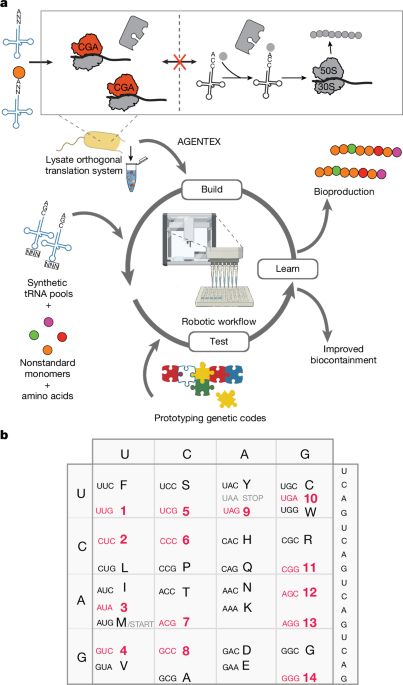
All protocols were run using the Jupyter notebook of OT-2. The protocol was divided into dsDNA production, DNA purification, tRNA production, tRNA purification, polypeptide production and polypeptide purification steps (polypeptide purification was optional and used for LC–MS/MS analysis but not for luminescence experiments). For all purifications, we specified the number of columns to be purified at the beginning of the protocol, and the multichannel pipette was used to carry out the procedure.
The tRNA pool sequences were automatically generated depending on the specified number of codons to be used for translation. The peptide DNA sequence was also generated with compressed genetic code from a provided amino acid sequence. All DNA sequences were ordered as chip-synthesized oligonucleotides and amplified and purified by machine protocols. All of the tRNAs in each tRNA pool were transcribed in one reaction, and all pools were purified in parallel. When an orthogonal aaRS, as in pazFRS used for M. jannaschii tRNAtyrCUA, was required, its dsDNA was also included in the translation reaction mixture. All reaction steps (dsDNA production, tRNA production, cell-free translation reaction) were inherited from a mixing script that separated sample data representation from machine control. Three .xlsx spreadsheets were provided containing layouts of two source plates as well as reactions. A .csv file was automatically generated that specified all operations the instrument would perform. The machine control protocol would then control the hardware and execute all steps.
Proteomic analysis of peptides produced in cell-free translation systems
Quantitative analysis was performed by a Q Exactive Orbitrap mass spectrometer (Thermo) equipped with an Evosep (Odense) nano-pump. The samples were run on 30 SPD (samples per day) method by Evosep. Flow-through that was size-filtered by 10 kDa filters was further digested in 50 mM TEAB buffer with trypsin for 3 h. The resulting digests were loaded directly onto Evotips (Evosep). The peptides were eluted from Evotips directly to 15 cm PepSep C18 (Bruker) column for chromatographic separation. The Q Exactive instrument was run in WWA mode (wide window isolation) with MS data-dependent acquisition. The data were searched by Thermo Proteome Discoverer 3.1.1.93 with Chimerys search engine. The data were searched against the general E. coli K12 database from Uniprot and a custom-built library database that included all predicted amino acid changes. We allowed variable modification on Ala (+4 Da) in cases in which isotopically labelled alanine was included in the cell-free translation system. All quantitation of the presence of such heavy amino acids was done based on MS spectra peak volumes that were automatically extracted by software from raw data. The results were kept at 0.1% FDR level on both peptide and protein level by Percolator64.
For analysis of AGENTEX crosstalk, quantitative analysis of pulldown samples from magnetic beads was performed with ZenoTOF7600+ (SCIEX) coupled with Evosep (Odense) nanoHPLC system. The samples were digested directly from the magnetic beads in 50 mM TEAB buffer at 37 °C over 3 h. The digested peptides were loaded directly into Evosep tips for analysis. The peptides were eluted from Evotips directly to 15 cm PepSep C18 (Bruker) column for chromatographic separation. The instrument was running in DIA mode with a 400–800 Da and automatic window isolation with 20 ms accumulation time for MS/MS acquisition. Searches were done with the PEAKS 13 database (BSI). All searches were done with 1 % FDR at protein and peptide levels. The data were searched against the general E. coli K12 database from Uniprot and a custom-built library database that included all predicted amino acid changes.
Reporting summary
Further information on research design is available in the Nature Portfolio Reporting Summary linked to this article.
Keep following us for the latest insights.